N-glycosylation at an evolutionary nexus

Echinoderms such as sea urchins and brittle stars are model organisms used to understand our own biology due to the evolutionary relationship. Two new papers from the Molecular Glycobiology group published in JBC on unusual glycans from this phylum reinforce the molecular similarities with vertebrates.

Two recent studies published in JBC, from the Molecular Glycobiology & Glycophylogeny group, headed by Iain Wilson, at the Department of Chemistry, address this gap in the knowledge of carbohydrate structures within these animals. The results obtained after purification using liquid chromatography and analysis using mass spectrometry and chemical and enzymatic treatments showed a wealth of hybrid and complex oligosaccharide structures reminiscent of those in higher vertebrates as well as some classical invertebrate glycan structures. The high degree of modification of the N-linked glycans with negatively charged groups, coupled with species-specific motifs, is a hint that sugar chains may be responsible for reproductive isolation and speciation. The studies also indicate the power of isomeric glycan analysis using off-line MALDI-TOF MS based on the accumulated experience of the postdoctoral FWF Fellows in the group.

Sulfated and sialylated N-glycans in the echinoderm Holothuria atra reflect its marine habitat and phylogeny
Jorick Vanbeselaere, Chunsheng Jin, Barbara Eckmair, Iain B. H. Wilson and Katharina Paschinger
J. Biol. Chem. 2020; 295:3159-3172 First Published On January 22, 2020. doi:10.1074/jbc.RA119.011701[Abstract][Full Text][Supporting Information][PDF]

Glycosylation at an evolutionary nexus: the brittle star Ophiactis savignyi expresses both vertebrate and invertebrate N-glycomic features
Barbara Eckmair, Chunsheng Jin, Niclas G. Karlsson, Daniel Abed-Navandi, Iain B. H. Wilson and Katharina Paschinger
J. Biol. Chem. 2020; 295:3173-3188 First Published On January 30, 2020. doi:10.1074/jbc.RA119.011703[Abstract][Full Text][Supporting Information][PDF]

New insights into the superfood of the honeybee

Royal jelly is necessary for the fertility and longevity of queen honeybees, but may also be bioactive in humans. Relevant molecular signals were revealed by in-depth glycomics as recently published by Dr. Alba Hykollari and Dr. Katharina Paschinger @molglyco in MCP and featured in ASBMB Today.

Royal jelly is widely believed to have health benefits, although the medical evidence is scarce. One thing the substance certainly does is promote caste development in honeybees, causing genetically identical larvae to develop differently. All bee larvae eat royal jelly secreted by worker bees for the first few days of life, but those picked out to be queens continue to feed on it until they pupate and beyond, whereas those that will become workers switch to honey and pollen. Biologists believe molecular signals in royal jelly drive larval bees to develop into queens, but the details of that signaling, including which molecule is most important and how it is recognized, were unclear.

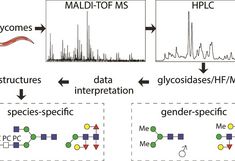
Questions along that line triggered Alba Hykollari and Katharina Paschinger, from the Molecular Glycobiology group at the Department of Chemistry, to revisit royal jelly. The results obtained after liquid chromatography and mass spectrometric analysis were surprising, since the glycosylation (a post translational modification of proteins) was more complicated and fascinating than expected from an insect. In this in-depth study around 100 different structures could be detected, especially complex and hybrid multiantennary N-glycans with fucose, sulfate, glucuronic acid and phosphoethanolamine. The recognition of phosphoethanolamine by a human immunoprotein has potential for anti-inflammatory effects on human health whereas the presence of fucoses can trigger allergic reactions.

Knowing these structures could help other scientists to understand the activity of glycosylated proteins in royal jelly and how they designate larval bees as future queens. The study was recently published in Molecular and Cellular Proteomics (MCP) and featured in ASBMB Today.

Link MCP: http://www.mcponline.org/content/17/11/2177

Link ASBMB: http://www.asbmb.org/asbmbtoday/201812/News/Honeybees/

The underestimated N-glycomes of lepidopteran species

A comparative N-glycomic study of the two lepidopteran (moth) pests revealed sulphate, glucuronic and zwitterionic N-glycan modifications. This is not only of evolutionary interest, but has biotechnological relevance as cell lines from these species are potential factories for recombinant glycoprotein production. Insects are significant to the environment, agriculture, health and biotechnology. Many of these aspects display some relationship to glycosylation. For a long time insect glycosylation was considered to be rather similar and simple, but more in-depth studies show structural diversity and interspecies variability.

In a new study, published in the April 2017 issue of ‘Biochimica et Biophysica Acta (BBA)‘, chromatographic and mass spectrometric methods were used to glycomically analyse two lepidopteran species (the cabbage looper Trichoplusia ni and the gypsy moth Lymantria dispar) as well as the commonly used T. ni High Five cell line. Thereby around 5% anionic N-glycan species, decorated with sulphate and glucuronic acid were detected, some also carrying the zwitterion phosphorylcholine. This degree of modification and variability is not only of evolutionary interest, but is of biotechnological relevance as lepidopteran cell lines are potential factories for recombinant glycoprotein production.

Link to the study: doi.org/10.1016/j.bbagen.2017.01.009

It’s proven: females are more complicated than males

Females are more complex than males, at least when it comes to N-glycosylation of the pig parasite Oesophagostomum dentatum. For the first time, gender-specific protein-linked oligosaccharide chains in a nematode worm could be revealed; thus, the synthesis of female-specific glycans requires more enzymatic processing steps than for the male ones.

Link to paper in Biochimica & Biophysica Acta