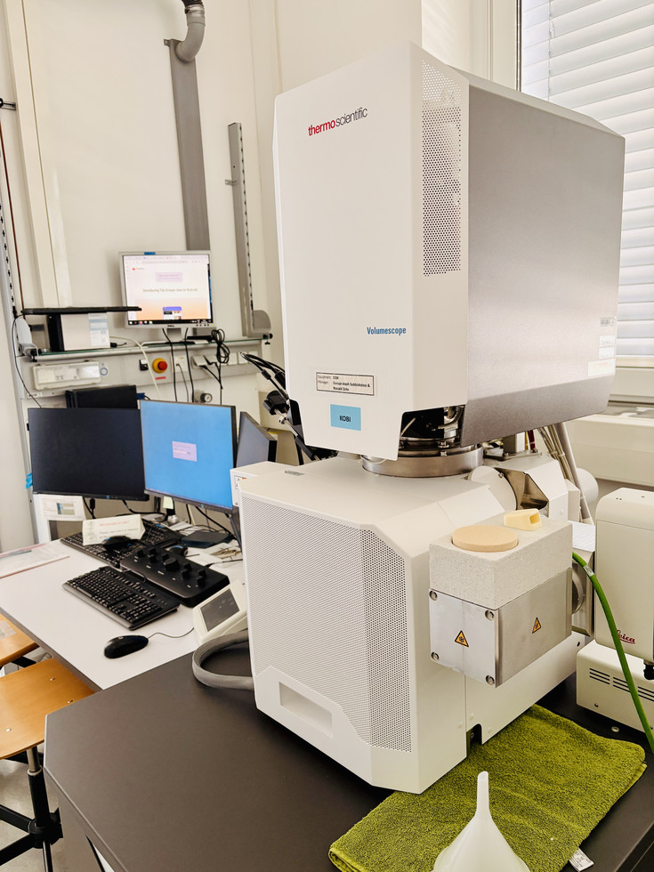
The FEI Apreo VolumeScope is a high-resolution field emission scanning electron microscope designed for advanced 2D and 3D imaging of biological, soft-matter, and materials-science samples.
A central capability of the instrument is serial block-face scanning electron microscopy (SBF-SEM). For SBF-SEM, resin-embedded samples are mounted inside the SEM and repeatedly imaged and sectioned using an integrated ultramicrotome. This automated image-and-cut workflow generates serial image stacks that can be used for 3D reconstruction of cells, tissues, plant material, microorganisms, biomaterials, and other structured specimens.
The Apreo VolumeScope can be used for room-temperature SEM imaging of prepared samples such as resin blocks, polished surfaces, coated specimens, fractured samples, and serial sections. It is particularly useful for biological ultrastructure, plant tissue analysis, cell and tissue morphology, biofilms, biomaterials, and nanobiotechnology applications.
The system also supports cryo-SEM workflows. In combination with our Leica cryo-preparation equipment, samples can be high-pressure frozen, freeze-fractured, coated, transferred under vacuum, and imaged under cryogenic conditions. This enables the analysis of frozen samples in a state closer to their native conformation and is especially valuable for hydrated, delicate, or soft biological specimens such as bacterial and mammalian cell cultures, plant material, biofilms, gels, and other sensitive samples.
In addition to block-face and cryo imaging, the instrument can be used to image array tomography samples, where serial sections are collected onto a suitable substrate and imaged in the SEM. This approach can complement SBF-SEM when physical serial sections need to be preserved, re-imaged, or combined with other imaging modalities.
The microscope is equipped with an EDS detector for energy-dispersive X-ray spectroscopy (EDS/EDX). EDS enables elemental analysis and mapping, providing compositional information in addition to structural SEM imaging. This can be useful for locating mineral deposits, metals, nanoparticles, stains, contrast agents, particles, or other inorganic components in biological and hybrid samples.
Image data from SEM, SBF-SEM, and array tomography workflows can be further processed and visualized using image analysis software such as Amira, which is available on the support workstation. Depending on the sample and acquisition strategy, this can support segmentation, 3D visualization, and quantitative analysis of volume datasets.