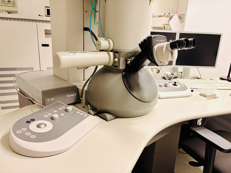
The FEI Tecnai G2 20 is a transmission electron microscope used for high-resolution ultrastructural analysis of biological samples and nanomaterials. The instrument was purchased in 2010 and complements the capabilities of the facility’s scanning electron microscopy platform by providing transmission electron microscopy imaging of ultrathin sections, negatively stained samples, particles, grids, and suitable electron-transparent specimens.
The TEM is particularly suited for the detailed analysis of cells, tissues, organelles, microorganisms, plant material, nanoparticles, nanostructured materials, and biomaterials. In biological applications, it enables visualization of intracellular ultrastructure, membranes, cell walls, organelles, viruses, bacteria, macromolecular assemblies, and other fine structural details that require transmission electron microscopy resolution.
The instrument can also be used for immunolocalization studies, for example immunogold labeling of specific proteins or cellular components on suitable sections or grids. This allows structural information to be combined with localization of defined molecular targets.
In addition to conventional TEM imaging, the Tecnai G2 20 supports electron tomography workflows for volumetric characterization of biological samples. By acquiring tilt series from suitable sections or specimens, three-dimensional reconstructions of selected sample volumes can be generated. This is useful for studying complex cellular architecture, membrane systems, organelles, interfaces, and nanoscale organization in biological or nanobiotechnology samples.
The use of the TEM is booked, tracked, and charged through the facility management software PPMS. After successful training, users typically operate the instrument independently. Assisted sessions can be requested through PPMS when additional support is required.
At present, sample preparation for TEM is primarily the responsibility of the users. This includes workflows such as ultrathin sectioning, negative staining, immunolabeling, cryo-fixation followed by freeze substitution, resin embedding, and grid preparation. The facility can provide guidance and, where appropriate, refer users to experienced personnel and established protocols, for example within the Institute of Plant Biotechnology and Cell Biology (IPBT).