Understanding heavy metal transport, tolerance, accumulation and adsorption dynamics in mosses
SUPERVISOR: Helga LICHTENEGGER
PROJECT ASSIGNED TO: Matthias WEINBERGER
Mosses are frequently used as biomonitors to investigate environmental pollution because of their ability to absorb both essential minerals and water, as well as harmful heavy metals, over their entire surface and their rhizoids. In this project we are mainly working with the moss species Physcomitrium patens and Pohlia drummondii. P. patens is often used as a model organism for non-vascular plants while P. drummondii is known to be a particularly heavy metal-resistant moss. This may be due to its thicker cell wall compared to P. patens. Mosses lack true vascular tissue which limits their height and many moss leaves consist of only a single layer of cells. These characteristics make them ideal candidates for imaging techniques and for analyzing metal transport and accumulation mechanisms in plants. Moss cell walls and cell walls of crop plants are composed of the same polysaccharides and are therefore comparable. Our findings could therefore lead to a potential model for heavy metal uptake in plants. To have better control over the applied metal pollution our moss samples are grown under controlled conditions in the laboratory of our project partners at the Department of Functional and Evolutionary Ecology, University of Vienna.
Due to the dimensions of a moss cell in the range of 20-60 µm and possible changes in ultrastructural features due to heavy metal contamination of only a few 100 nm, the methods used in this project focus on synchrotron X-ray applications, as only these light sources can provide the necessary spatial resolution. The techniques involved are X-ray fluorescence (XRF) mapping, X-ray absorption near edge structure (XANES) spectroscopy, extended X-ray absorption fine structure (EXAFS) spectroscopy and small and wide-angle X-ray scattering (SAXS/WAXS). There are also plans to combine these measurements with Raman spectroscopy to gain further insight into the composition of the moss cell wall.
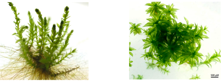
Figure 1: Habitus of the investigated moss species. (a) P. drummondii and (b) P. patens. Figure taken from Schillaci et al. Plants 2023, 12, 3960. doi.org/10.3390/plants12233960

Figure 2: XRF scan of a moss leaf contaminated by MnCl2: (a) shows the stereo microscope image of the leaf, (b) the potassium fluorescence signal, (c) the manganese fluorescence signal and (d) the Compton scattering ROI.